|
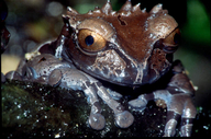
Triprion spinosus (Steindachner, 1864)
Coronated Treefrog | family: Hylidae subfamily: Hylinae genus: Triprion |
 © 2006 Dr. Peter Janzen (1 of 41) |
|
|
|
Description Distribution and Habitat Country distribution from AmphibiaWeb's database: Costa Rica, Honduras, Mexico, Panama
Life History, Abundance, Activity, and Special Behaviors Larva Tadpoles have been found in bromeliads on felled trees (Taylor 1954), water-filled tree cavities (Robinson 1961; Duellman 1970), and bamboo internodes in a botanical garden forest (Jungfer 1996). Jungfer (1996) reported on breeding in captivity. Eggs were laid into a treehole during daytime, over a period of 2 to 4 hours, although Jungfer (1996) considered the diurnal amplexus likely to be an artifact of captivity. An amplectant pair enters the treehole head down in the water and with the vents of the pair above water level. Four to five egg-laying bouts take place, each resulting in 3-5 fertilized eggs attached to the wall of the container just above the surface of the water. After egg deposition, the female leave the container; the male called softly a few times and leaves soon after. On subsequent nights, the male returns and mates again with the same female or occupies another container and starts calling again. In captivity, a clutch consisted on average of 158 eggs (range 48-311) with each egg having a diameter of 1.5 to 1.8 mm; eggs had a dark gray animal pole and a small white vegetal pole and were surrounded by a jelly coat. Only a few eggs (1-25) per clutch, 6% of total eggs, were fertilized and hatched in captivity. Larvae hatched after 6-7 days (Jungfer 1996). In captivity, the female first returned to the water-filled container about 7 days (range 5-9 days) after depositing fertilized eggs. Sometimes the male was present (resulting in oviposition as described above). If the male was not present, the female sat in the water with her cloaca submerged and laid up to 8 unfertilized eggs into the water, with the eggs mostly being grabbed and consumed by a tadpole as soon as they were extruded from the female's cloaca. Tadpole feeding occurred at intervals of about 5 days (range 1-14 days), for a total of 13-31 visits. Oviposition of the nutritive eggs took place only after the female received tactile stimuli from the tadpoles (swimming slowly around the mother, touching her with their mouths and sucking slightly at her skin, with movements becoming faster just before nutritive eggs were extruded). If a second clutch of fertilized eggs was laid, the subsequent larvae disappeared within two days, presumably eaten by their older siblings. After 60-132 days, 1-16 larvae metamorphosed from captive-bred clutches. The froglet is 26-28 mm. Larvae are able to breath atmospheric oxygen after hatching (Jungfer 1996). Trends and Threats The major threats to this species appear to be habitat loss and degradation, arising from smallholder farming and subsistence wood gathering (Santos-Barrera et al. 2004). Possible reasons for amphibian decline General habitat alteration and loss Comments This species was featured in News of the Week April 13, 2020: While frogs are well known for having skeletons that are reduced with respect to other terrestrial vertebrates, the skeleton still exhibits substantial variation among frogs, especially in the skull. Using three-dimensional anatomical data, Paluh et al. (2020) quantified frog skull diversity across all living families to test for relationships among ecology, skull shape, and increased mineralization (known as hyperossification or dermal ornamentation). Hyperossification has evolved more than 25 times across the frog tree of life, and although it can be present in species of nearly all sizes and habitats, it often occurs in frogs with deviant skull shapes that are known to either prey on other vertebrates or use their head to fill cavities or block holes (a behavior called phragmosis). Hyperossification is present in some frogs not known to feed on large prey or use phragmotic behavior, and it is possible that the function of hyperossification in these species may be tied to osmoregulation. Frogs are often assumed to share a highly conserved skeleton, but the study illustrates substantial diversity across anuran skulls that is linked to varied functions. (Dan Paluh) This species was featured in News of the Week August 5, 2024: In recent decades, large-scale digitization efforts have mobilized natural history collections making them central to transformative, modern research. The openVertebrate (oVert) Thematic Collections Network is one such innovative, large-scale digitization effort started in 2017, which focused on making 3D data of species available to a broad audience of scientists, students, teachers, artists, and more (Blackburn et al 2024). The high-fidelity 3D products include computed tomography (CT), diffusible iodine-based contrast-enhanced CT (diceCT), and photogrammetry images that are freely available for downloading from Morphosource, an online repository of high resolution biological images. The aim was not to scan every vertebrate species but rather provide a catalyst for launching museum collections as a resource for these modern technologies. Currently oVert scans are used in 3D reconstructions to visualize the bony and soft tissues for more than 13,000 vertebrate specimens. Although funding is officially complete, oVert has spawned over eight more NSF-funded CT digitization efforts with more indepth focus on everything from dinosaurs to Mesoamerican herpetofauna. The oVert project’s impact on amphibian research has been steadily growing with 96% of amphibian genera (548) scanned and available for use. So far over 413 scans of amphibian species have been used in a dozen scientific studies with more in progress. (Michelle Koo)
References
Duellman, W.E. (1970). The Hylid Frogs of Middle America. Monograph of the Museum of Natural History, University of Kansas. Jungfer, K.-H. (1996). ''Reproduction and parental care of the coronated treefrog, Anotheca spinosa.'' Herpetologica, 52(1), 25-32. McCranie, J. R., and Wilson, L. D. (2002). ''The Amphibians of Honduras.'' Contributions to Herpetology, Vol 19. K. Adler and T. D. Perry, eds., Society for the Study of Amphibians and Reptiles, Ithaca, New York. Robinson, D. C. (1961). ''The identity of the tadpole of Anotheca coronata (Stejneger).'' Copeia, 1961, 495. Santos-Barrera, G., Flores-Villela, O., Solís, F., Ibáñez, R., Savage, J., Chaves, G., and Kubicki, B. (2004). Anotheca spinosa. In: IUCN 2009. IUCN Red List of Threatened Species. Version 2009.1. www.iucnredlist.org. Downloaded on 07 September 2009. Savage, J. M. (2002). The Amphibians and Reptiles of Costa Rica:a herpetofauna between two continents, between two seas. University of Chicago Press, Chicago, Illinois, USA and London. Sessions, S. K. (1978). ''The chromosomes of Anotheca spinosa (Stejneger), family Hylidae.'' Herpetologica, 34, 70-73. Taylor, E. H. (1954). ''Frog-egg eating tadpoles of Anotheca coronata (Stejneger) (Salientia, Hylidae) .'' University of Kansas Science Bulletin, 36, 580-595. Originally submitted by: Peter Janzen (first posted 2005-06-30) Life history by: Michelle S. Koo (updated 2024-08-11)
Comments by: Michelle S. Koo (updated 2024-08-11)
Edited by: Kellie Whittaker, Michelle S. Koo (2024-08-11) Species Account Citation: AmphibiaWeb 2024 Triprion spinosus: Coronated Treefrog <https://amphibiaweb.org/species/673> University of California, Berkeley, CA, USA. Accessed May 23, 2025.
Feedback or comments about this page.
Citation: AmphibiaWeb. 2025. <https://amphibiaweb.org> University of California, Berkeley, CA, USA. Accessed 23 May 2025. AmphibiaWeb's policy on data use. |


 Map of Life