|
Phyllobates terribilis Myers, Daly & Malkin, 1978
Golden Poison Frog | family: Dendrobatidae subfamily: Dendrobatinae genus: Phyllobates |
| Species Description: Myers CW, Daly JW, and Malkin B. 1978. ''A dangerously toxic new frog (Phyllobates) used by Emberá Indians of Western Colombia, with discussion of blowgun fabrication and dart poisoning.'' Bulletin of the American Museum of Natural History, 161, 307-366. | |
 © 2009 Victor Fabio Luna-Mora (1 of 26) |
|
|
|
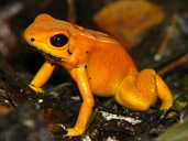
Description Phyllobates terribilis is a small frog (although large for a dendrobatid), with adult females having a maximum snout-vent length of 47 mm, and adult males reaching 45 mm in snout-vent length. Males mature at 37 mm while females mature at 40-41 mm. The snout is sloping and rounded in lateral profile, and bluntly rounded to truncate in dorsal view. The canthus rostralis is rounded, and the loreal region is both vertical and slightly concave. The tympanum is concealed posterodorsally. Hands and feet lack webbing and fringe. Fingers and toes terminate in moderately expanded discs; the disc of the third finger is expanded to the same degree in both males and females. If measured while appressed, the third finger is longest with the fourth, first, and second following in decreasing or approximately equal length; if measured from the base, the third finger is longest and the first finger is next while fingers 2 and 4 are roughly equal. Hands have a large, low, rounded tubercle on the palm. An inner metacarpal tubercle is present on the base of the first finger, with one subarticular tubercle on each of fingers 1 and 2, and two subarticular tubercles on fingers 3 and 4. On the feet, the relative length of the toes is 4>3>5>2>1. The distal third of the tarsus is ridged, with the ridge continuous from the inner metarsal tubercle to the tarsal tubercle. Toes 1 and 2 each bear one subarticular tubercle, toes 3 and 5 have two subarticular tubercles, and toe 4 bears three subarticular tubercles. Phyllobates terribilis skin is smooth to somewhat rugose or finely granular, becoming noticeably rugose and coarsely granular on the upper hind limbs. Teeth are present on the maxillary arch. Males have a shallow subgular vocal sac that is indicated by small expansion wrinkles at the base of the throat, and well-developed paired vocal slits on the floor of the mouth (Myers et al. 1978). The dorsal coloration on adult specimens of Phyllobates terribilis is bright golden yellow or golden orange, or pale metallic green, depending on the location of collection. Occasional individuals were deep orange or pale greenish yellow. Ventrally the color is the same as or slightly lighter than the dorsal color, except for the underside of the hands and feet, which are black, and the undersides of the thighs, which have a black seat patch. There is also a black crease at the axilla and groin. The eyes, nares and digit tips are black. There is usually black edging on the lower rim of the tympanum. In many individuals the mouth is edged with black, and the creases of limb articulations are often black. Males generally have some gray coloration on the base of the throat. In some individuals there is also a light gray suffusing the axilla and groin, as well as the posterior of the venter and the concealed part of the shank. In individuals that are pale metallic green in overall color, the venter may be slightly bluish green and the concealed part of the shank is sometimes a definite blue-green. Variation in ground color hue was associated with microgeographic variation (i.e. frogs collected from a given ridge or slope area tended to be the same hue) (Myers et al. 1978). Phyllobates terribilis tadpoles measure an average of 4.1 mm in body length and 11.1 mm in total length at hatching. At stage 37 (with well-developed hind legs), tadpoles have reached an average size of 12.6 mm in body length and 35.4 mm in total length. The head and body are depressed, with body width being greater than body depth. Eyes and nostrils are dorsal, and the eyes are directed dorsolaterally. The spiracle is sinistral and the vent is dextral (in some stage 25 larvae, the vent was located medially). The mouth is anteroventral with finely serrated beaks and 2/3 tooth formula; the second row of teeth above the mouth has a gap above the beak. The oral disc is anteriorly nude and indented laterally, with the lateral and posterior edges of the disc having one or two rows of papillae, depending on the developmental stage of the larva. Mouthparts are incompletely developed at stage 25 (the stage at which many dendrobatid tadpoles mount onto the back of the attendant parent frog). At stage 25 the denticles begin to keratinize and beak serrations are only just beginning to develop. Stage 25 larvae have a single row of papillae while stage 27 and older larvae have two rows (similar to the ontogenetic changes seen in tadpole oral papillae of some hylid frogs) (Myers et al. 1978). Newly hatched tadpoles are gray on the bodies and throats with paler gray tails and tail fins, with tiny flecks on the body and tail. At stage 26 the tail fins begin to become essentially transparent; there is weak pigmentation along the base of the fins and sparse flecking. At stage 37 the body color changes to blackish gray. Dense flecks are present dorsally and concentrate into paired dorsolateral stripes that run from the snout over the eyes to the base of the tail. To the naked eye the dorsolateral stripes look gray on a grayish black body, but under magnification the stripes appear pale bronze. At stage 42 (when forelimbs appear) the body is black and the dorsolateral stripes are brighter bronze to bronze-gold (Myers et al. 1978). Juvenile frogs are black in color, with gold dorsolateral stripes. This species undergoes an ontogenetic color change. As the frogs reach maturity, the dorsolateral stripes disappear, and the body becomes a more brightly colored uniform golden yellow. The bright golden dorsal coloration is achieved by 18 weeks, when the frog is about 21 mm in SVL, with the venter taking another several weeks to reach the same bright color. Since the frog reaches adult size in somewhat more than a year, the adult coloration is actually attained fairly early. Juvenile P. terribilis resemble P. aurotaenia in that both are black with paired gold dorsolateral stripes. However, juvenile P. terribilis can be distinguished by the lack of blue or green ventral spotting (present in juvenile P. aurotaenia) (Myers et al. 1978). The juvenile pattern of light stripes on a dark background is also lost in P. bicolor upon reaching maturity, but is retained into adulthood in the other members of the P. bicolor group, P. aurotaenia, P. lugubris and P. vittatus (Myers et al. 1978; Silverstone 1976). Distribution and Habitat Country distribution from AmphibiaWeb's database: Colombia
Life History, Abundance, Activity, and Special Behaviors Their calls are long sustained trills, consisting of a rapid succession of individual notes uttered at a rate of 13 per second, with a dominant frequency of 1.8 kHz. This frequency is lower than that of P. aurotaenia, P. bicolor, P.lugubris and P. vittatus (Myers et al. 1978). Eggs are laid terrestrially in small clutches of less than 20. Larvae, at developmental stage 25, are transported to pools on the backs of the male frogs. Male frogs have been observed carrying up to nine larvae simultaneously (Myers et al. 1978). The populations appear to be large and dispersed through the forest, with both adults and juveniles found on the forest floor, although juveniles appear to be relatively rare (2.5% of all specimens collected). Recruitment appears to be low since clutch size is small and few nurse frogs were observed carrying tadpoles, yet P. terribilis adults were fairly abundant at the type locality. This species takes more than a year to attain sexual maturity and is relatively long-lived (five years in captivity) (Myers et al. 1978). Given its toxicity, Phyllobates terribilis seems likely to have few predators other than the snake Leimadophis epinephelus. This snake has a high resistance to various anuran toxins (zetekitoxin from Atelopus zeteki, atelopid toxins from Atelopus elegans, piperidine alkaloids from Dendrobates auratus, and batrachotoxins from Phyllobates sp.). It is not a large snake (500 mm in total length); since adult Phyllobates terribilis frogs are fairly stocky and quite toxic, these snakes may only consume the juvenile P. terribilis and not the adults (Myers et al. 1978). During feeding in captivity, frogs may clasp each other around the head (cephalic clasping) or sometimes the body. Interestingly, Phyllobates terribilis males have been observed in captive aggressive behavior to press the upper surfaces of their hands against their opponent's chin (rather than grasping the other frog's head with the palms contacting the head) (Myers et al. 1978). Trends and Threats Relation to Humans Possible reasons for amphibian decline General habitat alteration and loss Comments This species produces the steroidal alkaloids batrachotoxin, homobatrachotoxin, and batrachotoxinin A. These compounds are extremely potent modulators of voltage-gated sodium channels, acting to keep the channels open and depolarizing nerve and muscle cells irreversibly, potentially leading to arrhythmias, fibrillation, and eventually cardiac failure (Albuquerque and Daly 1977). When accidentally transferred onto human facial skin, these toxins have been reported to cause a burning sensation lasting several hours (Myers et al. 1978). Batrachotoxin by itself is also capable of inducing numbness (see below for Myers' 1978 description of the effect of tasting P. vittatus), through its profound effects on a specific voltage-gated sodium channel (Na 1.8) known to be involved in pain reception. Thus batrachotoxin might be useful to develop as a topical pain-relief medication (Bosmans et al. 2004). The combination of batrachotoxin and homobatrachotoxin is produced in quantities up to 1900 micrograms per frog, which is at least 20-fold more than other toxic species in the family Dendrobatidae. The range of batrachotoxin-homobatrachotoxin produced by individual frogs was 700-1900 micrograms, with an average of 1100 micrograms per frog. The lethal dose of batrachotoxin-homobatrachotoxin for a 20 gram white laboratory mouse is .05 micrograms when injected subcutaneously. Thus one P. terribilis frog skin contains enough toxin to kill about 22,000 mice. The lethal dose of batrachotoxin for humans is not known but has been estimated at 200 micrograms, with a single frog thus potentially holding enough poison to kill about 10 humans. Wild-caught frogs that had been held in captivity for periods ranging from three weeks up to one year had toxicities of about 50% of recently caught specimens, with frogs maintained in captivity for three years still maintaining toxicity at about 40% the level of recently captured animals (Myers et al. 1978). In contrast, poison frogs born and reared in captivity do not contain toxins in their skin, although they can accumulate alkaloid toxins if these are part of the diet (Daly et al. 2004). Tadpoles did not contain batrachotoxin but a juvenile of 27 mm SVL was found to contain 200 micrograms of toxin, implying that the batrachotoxin alkaloids are synthesized or sequestered after metamorphosis (Myers et al. 1978). The toxins are concentrated in granular glands, which are most dense on the dorsal skin surfaces of the frog (Dumbacher et al. 2000). In contrast, the related species P. bicolor is much less toxic, containing from 17-56 micrograms of batrachotoxin-homobatrachotoxin per frog, with an average of 47 micrograms per individual. Despite P. bicolor being more brightly colored, larger, and more openly active than P. aurotaenia, the two species are thought to be roughly the same in toxicity levels (Myers et al. 1978). The more distantly related Central American species Phyllobates vittatus and P. lugubris also produce batrachotoxins but at much lower quantities. Despite these lower levels of toxin, Myers et al. (1978) reported that tasting the back of P. vittatus gave the human taster a sensation of near-numbness of the tongue, followed by a distinctly unpleasant tightening in the throat. Interestingly, batrachotoxins have also been found in feathers, but not the skin, of multiple species of passerine birds from New Guinea (particularly Pitohui dichrous, P. kirhocephalus, and Ifrita kowaldi) (Dumbacher et al. 2000). Breast, belly, and leg feathers show the highest toxin concentrations. Both toxin levels and toxin profiles in these birds vary considerably from population to population and can also change seasonally, implying that these toxins are acquired or synthesized from an environmental (dietary) source (Dumbacher et al. 2000). This source is most likely beetles of the genus Choresine (family Melyridae), which contain batrachotoxins and have been found in the stomach contents of New Guinea's toxic passerine birds. The family Melyridae is cosmopolitan and thus beetles could potentially be the source of the Phyllobates toxin as well (Dumbacher et al. 2004). Dietary arthropods (formicine ants, myrmecine ants, and in Madagascar, ponerine ants, as well as the siphonotid millipede Rhinotus purpureus) are known to provide defensive alkaloids to other toxic dendrobatid and mantellid frogs (Clark et al. 2005). This species was featured as News of the Week on 27 September 2021: What do Pitohui birds and Phyllobates poison frogs have in common? They both sequester the potent neurotoxin called batrachotoxin – and presumably resist its lethal effects. Batrachotoxin binds to voltage-gated sodium channels, a group of proteins that are partly responsible for action potentials, and thus interrupt the nervous system functions. Previous studies have predicted that the voltage-gated sodium channels from Phyllobates poison frogs possess changes in the amino acid sequence that prevent batrachotoxin from binding– yet Abderemane-Ali et al. (2021) find that batrachotoxin readily binds to poison frog and pitohui sodium channels, suggesting that these animals have a different mechanism of toxin resistance. They propose that circulating "toxin sponges" could bind to batrachotoxin, preventing it from binding to the voltage-gated sodium channels. Although a batrachotoxin-binding protein remains elusive, a unique toxin-binding protein called saxiphilin (which binds saxitoxin, the toxin underlying red tides) was discovered in bullfrogs (Rana catesbeiana) in the 1990s, suggesting that toxin sponges may be a common way for frogs to counter the potential lethal effects of toxin accumulation. Perhaps even more intriguing is the potential for "toxin sponges" to allow toxin sequestration and resistance to evolve in parallel (Marquez 2021, Abderemane-Ali et al 2021). Written by RTarvin
References
Albuquerque, E. X. and Daly, J. W. (1977). ''Batrachotoxin, a selective probe for channels modulating sodium conductances in electrogenic membranes.'' The Specificity and Action of Animal, Bacterial and Plant Toxins. Receptors and Recognition, Series B., Volume 1 P. Cuatrecasas, eds., Chapman and Hall, London, 297-338. Bosmans, F., Maertensa, C., Verdonck, F., and Tytgat, J. (2004). ''The poison Dart frog’s batrachotoxin modulates Nav 1.8.'' Federation of European Biochemical Societies Letters, 577(2004), 245-248. Clark, V. C., Raxworthy, C. J., Rakotomalala, V., Sierwald, P., and Fisher, B. L. (2005). ''Convergent evolution of chemical defense in poison frogs and arthropod prey between Madagascar and the Neotropics.'' Proceedings of the National Academy of Sciences, 102(33), 11617-11622. Daly, J. W., Secunda, S. I., Garraffo, H. M., Spande, T. F., Wisnieski, A., and Cover, J. F. (1994). '' An uptake system for dietary alkaloids in poison frogs (Dendrobatidae).'' Toxicon, 32(6), 657-663. Dumbacher, J. P., Spande, T. F., and Daly, J. W. (2000). ''Batrachotoxin alkaloids from passerine birds: A second toxic bird genus (Ifrita kowaldi) from New Guinea.'' Proceedings of the National Academy of Sciences, 97, 12970-12975. Dumbacher, J.P., Wako, A., Derrickson, S.R., Samuelson, A., Spande, T.F., and Daly, J.W (2004). ''Melyrid beetles (Choresine): a putative source for the batrachotoxin alkaloids found in poison-dart frogs and toxic passerine birds.'' Proceedings of the National Academy of Sciences of the United States of America, 101, 15857–15860. Maxson, L. R., and Myers, C. W. (2007). ''Albumin evolution in tropical poison frogs (Dendrobatidae): a preliminary report.'' Biotropica, 17(1), 50-56. Myers, C. W., Daly, J. W., and Malkin, B. (1978). ''A dangerously toxic new frog (Phyllobates) used by Emberá Indians of Western Colombia, with discussion of blowgun fabrication and dart poisoning.'' Bulletin of the American Museum of Natural History, 161, 307-366. Originally submitted by: Kellie Whittaker and Shelly Lyser (first posted 2005-02-14) Edited by: Kellie Whittaker, Michelle S. Koo (2021-09-26) Species Account Citation: AmphibiaWeb 2021 Phyllobates terribilis: Golden Poison Frog <https://amphibiaweb.org/species/1707> University of California, Berkeley, CA, USA. Accessed Jan 15, 2025.
Feedback or comments about this page.
Citation: AmphibiaWeb. 2025. <https://amphibiaweb.org> University of California, Berkeley, CA, USA. Accessed 15 Jan 2025. AmphibiaWeb's policy on data use. |


 Map of Life